We’ve all heard the old adage, “No foot, no horse.” The foot of the horse is a unique structure that plays a vital function in the overall health of the horse. There are many factors that affect the horse’s foot, from conformation and genetics to farrier and nutrition. As a horse owner, having an understanding of hoof anatomy and the many components that affect its structure can help you identify, treat, and prevent problems.
Anatomy
Internal:
The horse foot comprises bones with synovial (joint) spaces between, supported by tendons, ligaments, and the laminae of the hoof wall. There are no muscles in the foot! The three bones are the coffin (aka “pedal”) bone, the pastern bone, and the navicular bone. The coffin bone essentially equates in humans to the last bone on a person’s middle finger. Imagine that one structure bearing all the weight of a horse, and you can see why proper alignment and support of the bones is so important!
The bone just above (in veterinary lingo, “proximal to”) the coffin bone is the short pastern bone, while the smaller bone adjacent and behind (“caudal”) to these two bones is the navicular bone. The pastern and coffin bone are weight-bearing bones of the foot, while the navicular mainly serves as a fulcrum for the deep digital flexor tendon that runs down the back of the leg and attaches to the back of the coffin bone. The space between the coffin and pastern bone is called the “coffin joint,” and may be a site for arthritis in horses that have excess concussion on their feet. The synovial space around the navicular bone is called the “navicular bursa.” The fluid in this bursa helps cushion the navicular bone and the deep digital flexor tendon. There are also many soft tissue structures that support the bones of the foot. These include the deep digital flexor that runs from near
the carpus (front limb) or hock (hind limb) to the coffin bone (blue); small ligaments that surround the navicular bone (green); the common digital extensor on the dorsal (front) side of the joint (purple); and two short collateral ligaments on either side of the coffin joint (brown). Any of these tendons or ligaments can become injured and cause lameness. Unfortunately, these are difficult to see on ultrasound, so MRI is often the best way to truly diagnose injuries in these structures.

External:
The external foot comprises the frog, sole, white line, hoof wall, coronary band, and heel bulbs. The frog is the heart-shaped structure on the bottom of the foot that is responsible for expanding as the foot contacts the ground. It is important in shock absorption and circulation. The outer wall and sole are made of insensitive keratin, much like human fingernails. This protective capsule interdigitates with “sensitive” laminae surroudning the coffin bone that are more flexible and have nerve and blood supply. The hoof wall grows down from the coronary band (where the hair meets the hoof wall) at a rate of approximately 0.25” per month, and joins the sole (bottom of the foot) at a juncture called the “white line.”
Trimming and Shoeing
One of the most common questions we are asked is, “Does my horse need shoes?” The answer is that it depends on the horse’s job, conformation, and any previous or current injuries. A horse in no or light work with overall good conformation may be just fine with good, balanced trimming, while a horse with abnormal conformation or in heavy work may need shoes for support and traction.
The most important aspect, whether shod or not, is that the foot is appropriately balanced for the horse. What this means is that the horse’s foot has a good alignment from pastern to bottom of the foot (“hoof-pastern axis”), a wide frog, adequate sole depth, and overall symmetry. Although there are proposed standard numbers and angles to define “balance,” the reality is that it will vary somewhat from horse to horse and even from one limb to another depending on conformation.
Poor or irregular trimming will contribute to uneven and increased workload on the limb. Some common examples are overlong toes, hoof wall flares or cracks, and underrun and/or contracted heels. Abnormalities will increase strain on the hoof and soft tissues, which can lead to increased rate of injury or development of problems such as hoof abscesses, thrush, and white line disease.
In addition to the traction and support that can be provided by regular shoes, corrective trimming
and special shoes can be used to assist in injury. Some examples of special shoes and techniques include wedging, bar shoes, or pads.
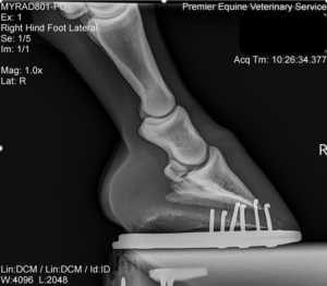
One useful way to assess trimming and relation to the structures inside the foot is by having foot x-rays performed (figures 5 and 6). Having a lateral (side-to-side) and dorsopalmar (front-to-back) view of each foot can reveal coffin bone imbalances or rotation, alignment of the coffin bone and pastern bones, sole depth, and joint disease (arthritis). This can be very useful in guiding trimming and shoeing decisions.


